
Erythrocyte morphology
Open each change to see what it looks like, why it happens and species traps.
Educational draft for review. Some photos come from openly licensed collections and are credited under each image.
Come to the lab
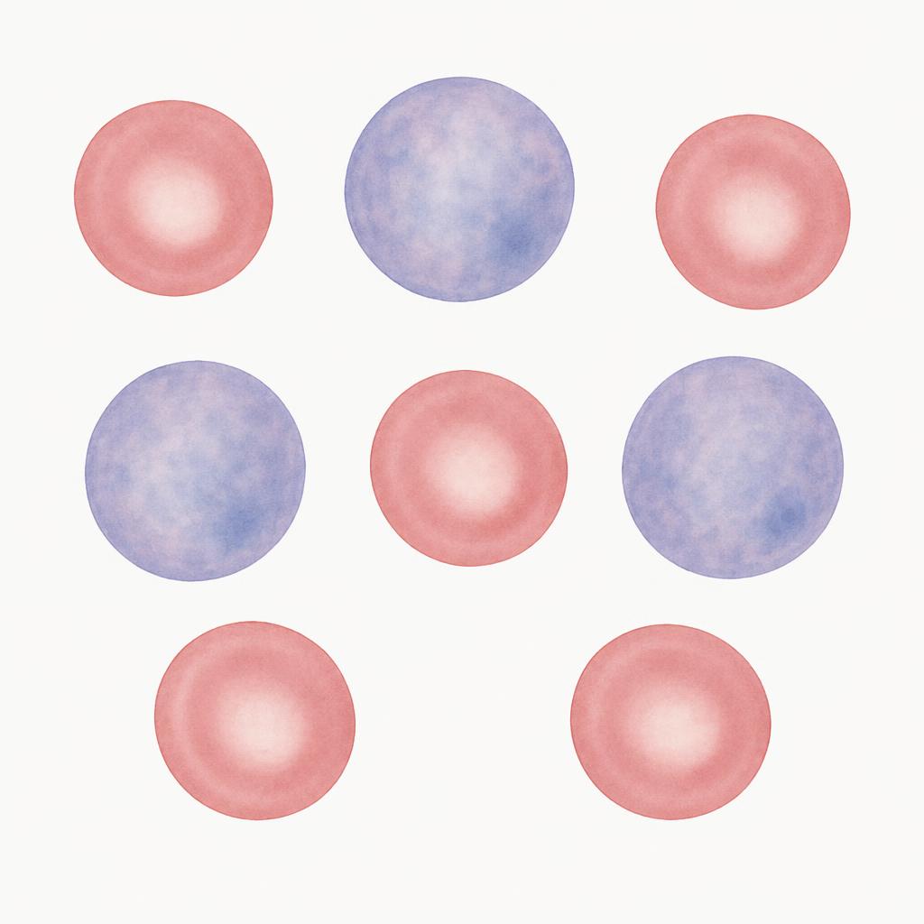
Polychromasia
What it looks like: Larger, blue-grey red cells mixed with mature pink cells.
What it means: Young red cells released by a responding marrow. Confirm with a reticulocyte count.
Species notes: Little or none in healthy horses, even when the marrow responds.
Spherocytes
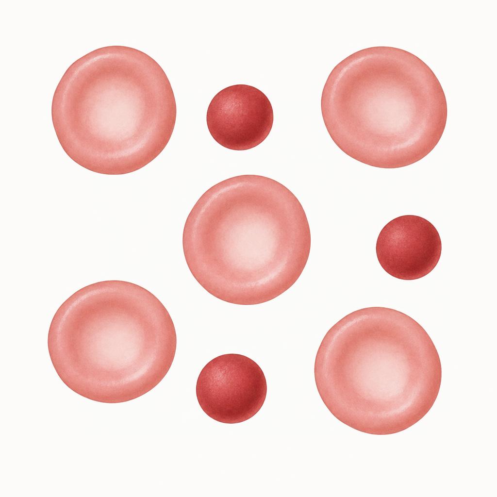
What it looks like: Small, round, dense red cells with no central pallor.
What it means: Partial removal of membrane by macrophages, most often immune-mediated hemolysis. Also seen after transfusion and some toxins.
Species notes: Easy to spot in dogs. Hard to judge in cats and horses because their cells lack obvious pallor.
Anisocytosis
What it looks like: Red cells of varying size.
What it means: Mixed populations: regeneration, iron deficiency, or recent transfusion.

Hypochromasia
What it looks like: Wide pale centre with a thin rim of colour.
What it means: Reduced hemoglobin per cell, classically chronic iron deficiency from blood loss. Do not confuse with bowl-shaped cells that only look pale.

Schistocytes
What it looks like: Small, irregular red cell fragments with sharp points.
What it means: Shearing in damaged vessels: DIC, hemangiosarcoma, vasculitis, heartworm disease, iron deficiency.

Acanthocytes
What it looks like: Few irregular blunt projections of unequal length.
What it means: Altered membrane lipids: liver disease, hemangiosarcoma in dogs, fragmentation disorders.

Echinocytes
What it looks like: Many short, evenly spaced, regular projections.
What it means: Usually a drying or storage artifact. True forms occur with electrolyte loss, kidney disease, snake envenomation.

Heinz bodies
What it looks like: Small round pale bumps on the cell edge; blue with new methylene blue stain.
What it means: Denatured hemoglobin from oxidants.
Species notes: Cats can have a few normally; many suggest oxidant exposure, diabetes or hyperthyroidism.
Eccentrocytes
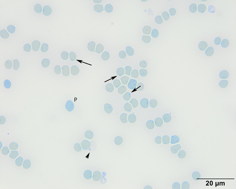
What it looks like: Hemoglobin is pushed to one side, leaving a clear crescent bounded by fused cell membrane.
What it means: Oxidative injury cross-links opposing membranes. Look for Heinz bodies and investigate oxidant exposure, including onions or garlic in dogs and red maple leaves in horses.
Keratocytes
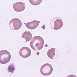
What it looks like: Cells with one or two horn-like projections around a blister.
What it means: Oxidative or mechanical injury, iron deficiency.

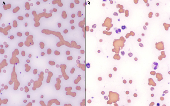
Agglutination
What it looks like: Grape-like clusters of red cells that persist after diluting with saline.
What it means: Antibody cross-linking cells, strongly supports immune-mediated hemolysis. Rouleaux disperse in saline; agglutination does not.
Species notes: Rouleaux is normal in horses and common in cats.
Howell-Jolly bodies
What it looks like: A single small dark round dot inside the cell.
What it means: Nuclear remnant. Seen with regeneration or after splenectomy.
Species notes: A few are normal in cats and horses.

Basophilic stippling
What it looks like: Fine blue dots throughout the cell.
What it means: Regeneration in ruminants and cats; lead poisoning in dogs when anemia is mild.

Red cell parasites
What it looks like: Small dots, rings or chains on or in the cell.
What it means: Mycoplasma (epicellular, on the surface), Babesia (paired teardrops inside), Anaplasma in cattle (dot on the edge). Do not confuse with stain precipitate.
