Leukocyte morphology
Start with granules and size, then open each cell to see what it looks like, why it changes and species traps.
Educational draft for review. Some photos come from openly licensed collections and are credited under each image.
Granules or no granules, and who is biggest
White cells split into two families by what is packed in the cytoplasm. The granulocytes (neutrophil, eosinophil, basophil) carry visible granule packets, which are tools the cell releases when it works. The agranulocytes (lymphocyte, monocyte) show no such packet on a routine smear.
| Cell | Cytoplasmic granules | What you actually see |
|---|---|---|
| Neutrophil | Yes, fine and pale | Pale pink-grey cytoplasm with dust-like granules that are easy to miss at low power. Toxic change makes them coarse and dark blue-purple. |
| Eosinophil | Yes, large and obvious | Brick-red to orange granules packed tightly through the cytoplasm. Their shape and colour change with species. |
| Basophil | Yes, coarse and dark | Deep purple to blue-black granules that often sit on top of the nucleus and hide it. |
| Monocyte | No visible specific granules | Grey-blue ground-glass cytoplasm, often with vacuoles and a faint dust, but no bright granule packet. |
| Lymphocyte | No visible specific granules | Clear to sky-blue cytoplasm with no granule packet. A few large granular lymphocytes carry a small number of red-purple granules, which is normal. |
Strictly speaking every white cell holds some primary granules. The names describe the specific granules you can see with a routine stain, which is why a monocyte counts as agranular even though it is not empty.
Cell size: typical ranges, not a fixed ranking
These comparisons use a dog red cell about 7 µm across and refer to diameter, not area. Many white cells really do overlap in size, so “about twice a red cell” cannot distinguish them. A 14 µm cell is twice the diameter but covers roughly four times the area of a 7 µm red cell. Species, smear preparation and activation also affect size.
| Cell | Typical diameter | Diameter relative to a 7 µm dog red cell | Why it matters |
|---|---|---|---|
| Monocyte | 15 to 20 µm | 2.1 to 2.9 × the diameter | Usually the largest normal circulating white cell; size overlaps with other large cells |
| Eosinophil | 13 to 15 µm in the dog | 1.9 to 2.1 × the diameter | Often slightly larger than a neutrophil, but their size ranges overlap |
| Basophil | 13 to 15 µm | 1.9 to 2.1 × the diameter | Similar in size to an eosinophil or large neutrophil; granules are more useful than size for identification |
| Neutrophil | 12 to 15 µm in the dog, 10 to 13 in the cat, 10 to 12 in cattle | Dog: 1.7 to 2.1 × the diameter | A mid-sized white cell; use red cells from the same species as your local ruler |
| Reactive lymphocyte | 12 to 15 µm, sometimes larger | 1.7 to 2.1 × the diameter at 12 to 15 µm | Can overlap with neutrophils or larger cells; deep blue cytoplasm helps identify it |
| Small lymphocyte | 7 to 10 µm | 1.0 to 1.4 × the diameter | Usually the smallest normal circulating white cell; about red-cell sized to slightly larger |
| Scale: dog red cell | About 7 µm | Your ruler | Use it to estimate anything sitting next to it |
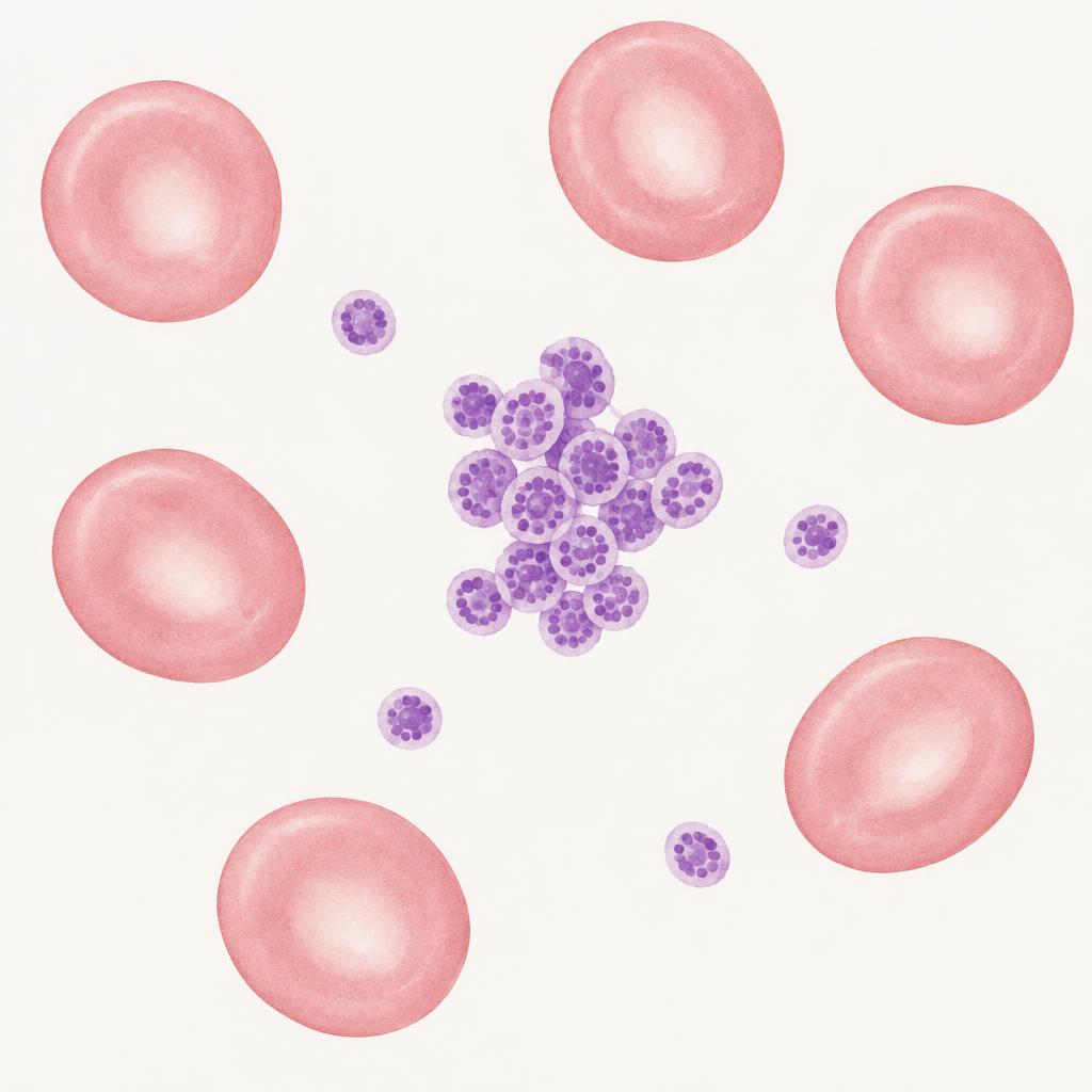
| Scale: platelet | 2 to 3 µm | About a third of a red cell | Smallest blood cell of all, and it has no nucleus |
The useful endpoints are the monocyte (usually largest) and small lymphocyte (usually smallest). Eosinophils, basophils, neutrophils and reactive lymphocytes overlap, so identify them by nucleus, cytoplasm and granules together, not size alone. Blasts vary in size; abnormal chromatin and nucleoli matter more than being the biggest cell.
Species traps
- Cat: eosinophil granules are small, rod or barbell shaped and often few in number, so a cat eosinophil can look almost empty.
- Greyhound: eosinophil granules are grey and vacuolated rather than orange.
- Horse: eosinophils are large with big round granules and are easy to mistake for another cell.
- Cattle and goat: neutrophils are smaller than a dog's and lymphocytes outnumber them, so most white cells on the smear look small.
- Bird and reptile: red cells keep their nucleus and are larger than the white cells, so the size order learned in mammals does not transfer.
Come to the lab
Segmented neutrophil
What it looks like: Pale cytoplasm, nucleus with three to five lobes.
What it means: Baseline cell. Increases with inflammation, stress and excitement.

Band neutrophil (left shift)
What it looks like: Horseshoe nucleus with parallel sides and no constrictions.
What it means: Marrow releasing immature cells because demand is high. A degenerative left shift (bands near or above mature cells with a normal or low count) signals overwhelming demand.
Species notes: Cattle shift left quickly because their marrow reserve is small.

Toxic change
What it looks like: Foamy or blue cytoplasm, Döhle bodies, toxic granules.
What it means: Rapid production during strong inflammation, often bacterial. Grade it from mild to severe.
Species notes: A few Döhle bodies can be normal in cats.
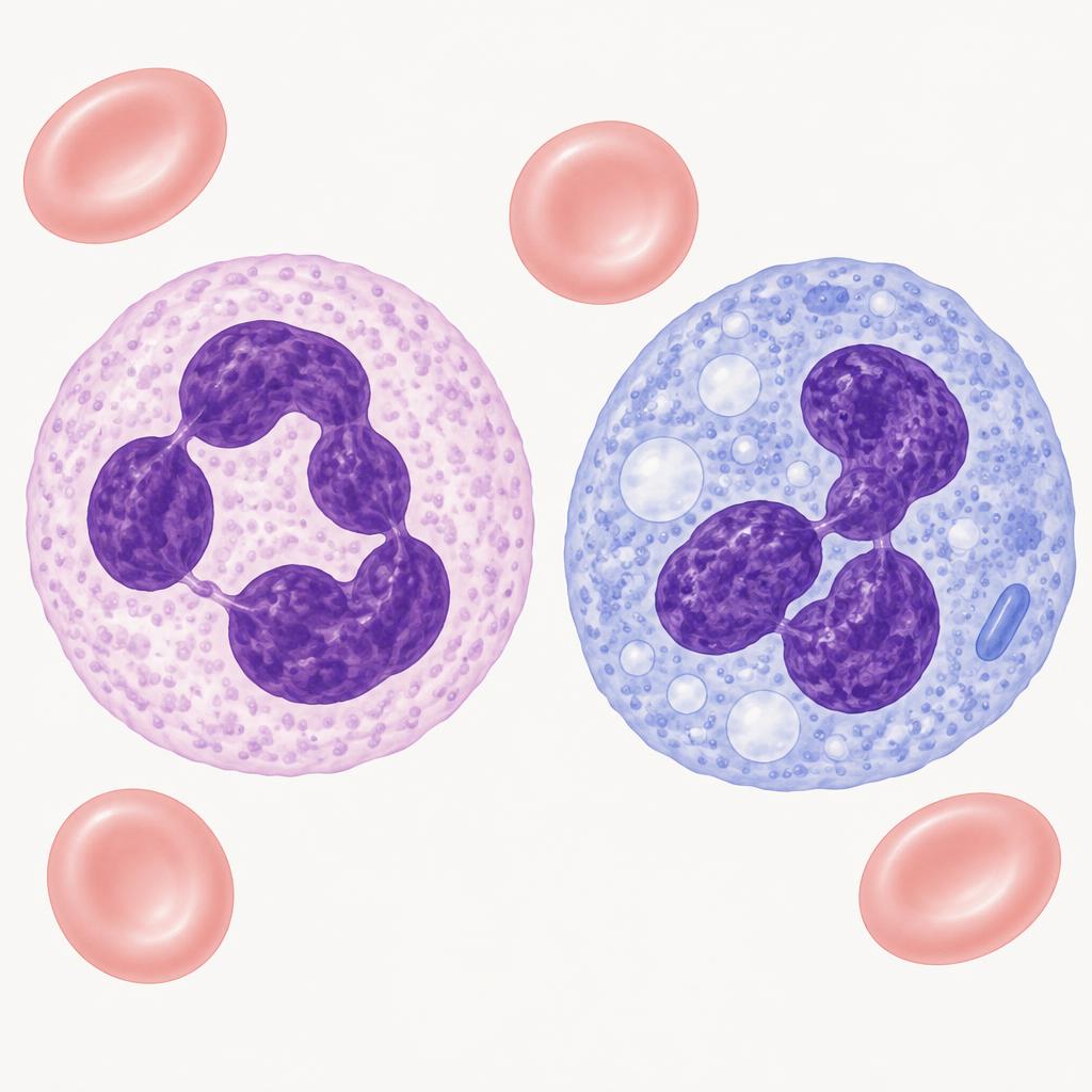
Hypersegmentation
What it looks like: Neutrophil with more than five lobes.
What it means: Aged cells staying longer in blood: steroids, stress, delayed sample processing.

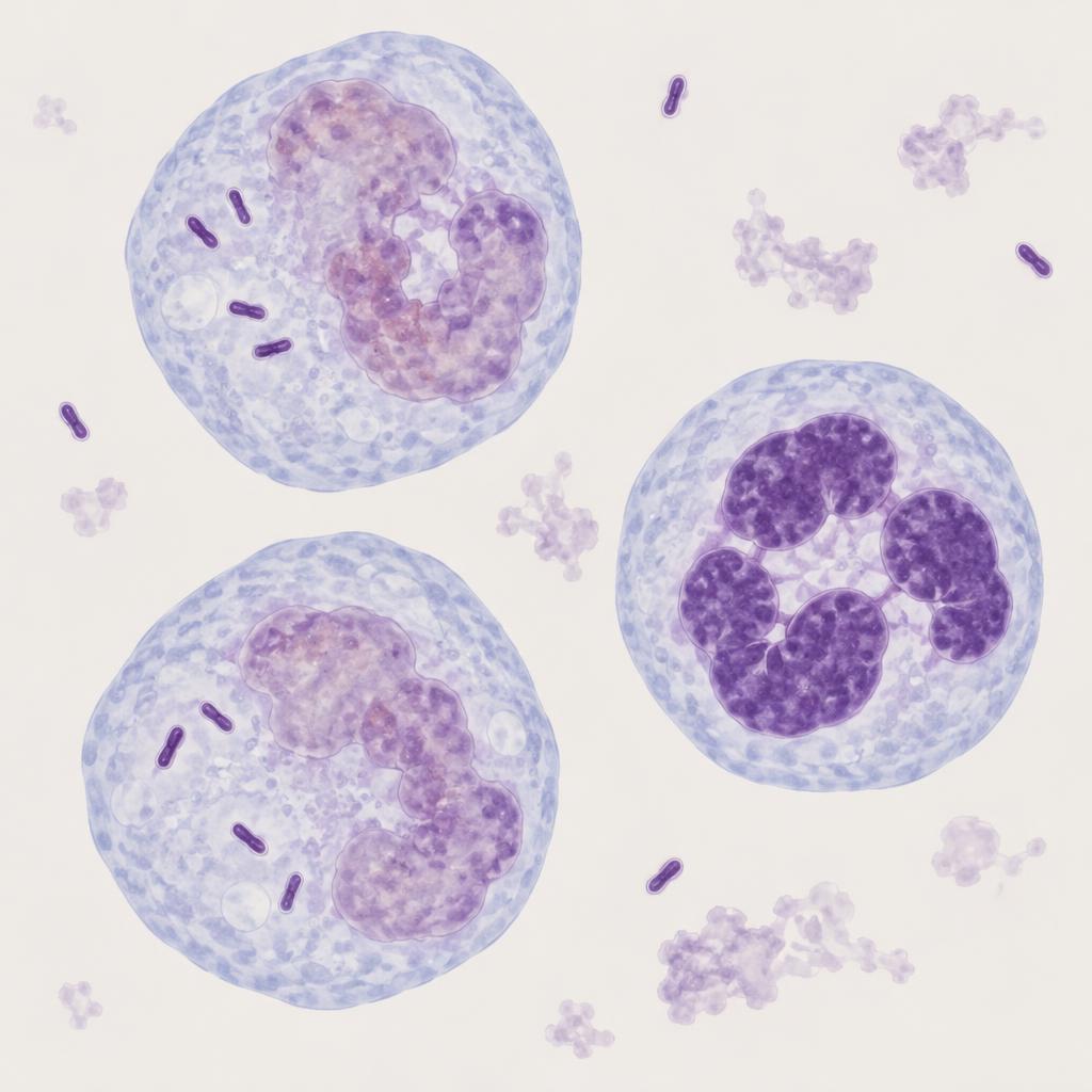
Intracellular bacteria
What it looks like: Bacteria inside intact neutrophils.
What it means: Sepsis or septic inflammation. Bacteria only outside cells may be contamination.
Lymphocytes: small and reactive
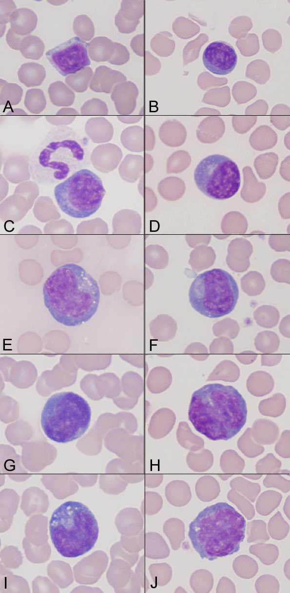
What it looks like: Small cells with scant blue cytoplasm; reactive cells are larger with deep blue cytoplasm.
What it means: Reactive forms suggest antigen stimulation. Persistent increases of medium to large cells raise concern for leukemia.
Monocytes
What it looks like: Large cells, variable nucleus, grey-blue cytoplasm, often with vacuoles.
What it means: Increase with chronic inflammation, necrosis and stress in dogs.

Eosinophils
What it looks like: Orange-red granules.
What it means: Parasites, allergy, some tumors such as mast cell tumors, hypoadrenocorticism (a normal count in a stressed sick dog is suspicious).
Species notes: Greyhounds have grey, vacuolated granules; cats have rod-shaped granules; horses have large round granules.

Basophils
What it looks like: Lavender to grey granules, often with a ribbon-like nucleus.
What it means: Usually alongside eosinophilia. Rare in dogs.

Blast cells
What it looks like: Large cells, fine chromatin, nucleoli, deep blue cytoplasm.
What it means: Should not circulate. Suggest acute leukemia or severe marrow disturbance; flow cytometry helps classify.

Platelet clumps
What it looks like: Aggregated platelets, often at the feathered edge.
What it means: Falsely low platelet counts. Always scan the feathered edge before calling thrombocytopenia.
Species notes: Very common in cats.